
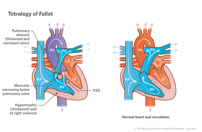
Tetralogy of Fallot (TOF) is a combination of defects of the heart. They include:
- Ventricular septal defect (VSD) - a hole in the wall of the heart between the pumping chambers of the heart (ventricles) which allows oxygen-rich “red” blood to pass from the left ventricle through the hole and mix with oxygen-poor “blue” blood in the right ventricle.
Mixing of the blood causes changes to oxygen levels in the body and the heart has to work harder to maintain the body’s requirements. It also causes an increase in blood flow to the lungs and may expose the lungs to high pressure. Learn more about ventricular septal defect.
- Pulmonary stenosis or right ventricular outflow tract obstruction – an obstruction to the flow of blood from the right ventricle to the pulmonary artery and lungs.
- Right ventricular hypertrophy – a thickening of the muscle in the right ventricle.
Overriding aorta – the aorta is wrongly positioned over both ventricles and receives mixed blood from both sides of the heart.
Signs and symptoms
The main signs and symptoms of VSD include:
- Blue skin, nails and lips
- Cool and clammy skin
- Irritability/tiredness/floppy body (but awake)
- Some children may have episodes of extreme blueness (hypercyanotic or ‘tet’ spells) in which they may become floppy, dazed or unconscious. These can be life-threatening and the child should be taken to the emergency department. Learn more about Tet spells.
Diagnosis
Tests to help diagnose Tetralogy of Fallot include:
- Electrocardiogram (ECG) – measures the electrical activity of the heart.
- Echocardiogram (Echo) – uses sounds waves to produce a moving picture of the heart.
- CT Scan – an X-ray with dye injected to highlight blood vessels.
- Cardiac catheterisation – A thin flexible tube (called a catheter) is inserted into a blood vessel in the groin and fed up into the heart to measure pressures and oxygen levels, and visualise heart structures using X-ray equipment. The procedure is performed under a general anaesthetic. Learn more about cardiac catheterisation.
These tests are usually carried out because a doctor has heard a heart murmur when examining a child with a stethoscope or detected during an pregnancy ultrasound.
Treatment
The treatment for Tetralogy of Fallot is surgery.
Surgery options include:
- using a patch to close the hole
- a shunt operation – a small tube is inserted between the aorta and the pulmonary arteries to help increase the amount of blood that can get to the lungs.
When to seek help
Call Triple Zero (000) and ask for an ambulance if your child:
- Looks pale or has blue lips, skin or nails.
- Is floppy, dazed or unconscious.
If it’s not an emergency but you have any concerns or questions, contact your GP or 13 Health (13 43 2584). If your child is under the care of a cardiologist, contact their cardiac care coordinator.
Developed by the Cardiology Department, Queensland Children's Hospital. We acknowledge the input of consumers and carers.
Resource ID: FS240. Reviewed: January 2024
Illustrations republished with permission from The Royal Children's Hospital, Melbourne, Australia. Images subject to copyright.
Disclaimer: This information has been produced by healthcare professionals as a guideline only and is intended to support, not replace, discussion with your child’s doctor or healthcare professionals. Information is updated regularly, so please check you are referring to the most recent version. Seek medical advice, as appropriate, for concerns regarding your child’s health.