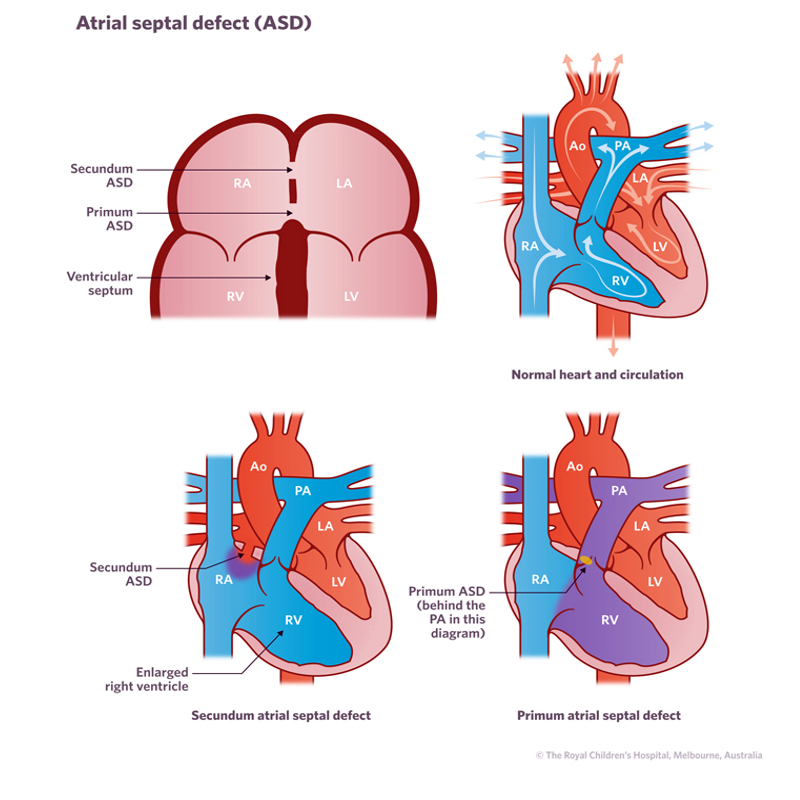
An atrial septal defect (ASD) is a heart condition in which there is a hole in the atrial septum (the wall that separates the left and right chambers (or atrium) of the heart. The atrial septum separates the oxygenated blood and the deoxygenated blood within the heart. A hole in the atrium septum allows blood to pass from one side of the heart to the other, usually from left to right. This can lead to the heart enlarging on the right side and lung circulation becoming overloaded, and can also cause abnormal heart rhythms. Atrial septal defects can vary in size and position.
The cause of atrial septal defect is unclear, but they typically occur while a baby is developing in the womb. Genetics, certain medical conditions, use of certain medications, and environmental or lifestyle factors, such as smoking or alcohol misuse, may play a role.
Atrial septal defects account for 5-10% of all cases of congenital heart disease and are twice as prevalent in girls as boys.
About half of all atrial septal defects close spontaneously without treatment.

Signs and symptoms
The main signs and symptoms of an atrial septal defect include:
- tiredness/fatigue with exertion
- chest infections, shortness of breath
- heart palpitation, irregular heartbeat
- poor growth in developing children
A large hole left untreated can lead to heart failure and heart arrhythmias later in life. However, most people are normally active and show no outward signs of illness.
Diagnosis
Tests that help diagnose atrial septal defect include:
- Electrocardiogram (ECG) – measures the electrical activity of the heart.
- Echocardiogram (Echo) – uses sound waves to produce a picture of the heart.
- Cardiac catheterisation – A thin, flexible tube (called a catheter) in inserted into a blood vessel in the groin and fed up into the heart to measure pressures and oxygen levels, and visualise heart structures using X-ray equipment. The procedure is performed under a general anaesthetic. Learn more about cardiac catheterisation.
- Transoesophageal echo - a flexible telescope with an ultrasound scanner attached to it placed down the oesophagus to provide close-up images of the heart.
- MRI or magnetic resonance imaging – non-invasive imaging procedure that produces detailed images of organs and structures in the body.
These tests are usually performed because a doctor has heard a heart murmur (turbulent blood flow) when examining a child with a stethoscope.
Treatment
Some atrial septal defects can be closed via cardiac catheterisation. Under a general anaesthetic a fine tube is inserted into a blood vessel in the groin and fed up to the heart chambers. The device is then passed through the catheter into the hole and expanded, acting like a plug in the hole. This plug remains in the septum and the catheter is removed. Eventually the tissue of the heart grows over the device making it part of the heart.
Some atrial septal defects have to be closed surgically. Under a general anaesthetic, the hole is closed with stitches or a patch (either tissue or synthetic).
When to seek help
See your GP or nearest hospital if your child has:
- Shortness of breath
- Easy tiring, especially after activity
- Swelling of legs, feet, or belly (abdomen)
- Sensations of a rapid, pounding heartbeat (palpitations) or skipped beats.
If it’s not an emergency but you have any concerns or questions, contact your GP or 13 Health (13 43 2584).
In an emergency, call Triple Zero (000) and ask for an ambulance.